Researchers seek to unlock details on developmental defects
LAWRENCE — Researchers at the University of Kansas School of Engineering have partnered with the University of Kansas Medical Center on a project to better understand developmental biology and how an embryo evolves during its earliest stages.
 “The goal is to eventually learn more about developmental defects,” said Prajna Dhar, assistant professor of chemical and petroleum engineering and one of the leaders on the project. “The more we know about embryo development and why it forms the way it does could help us to eventually unlock some of the reasons defects form.”
The project, which recently received a $1.2 million, four-year grant from the National Institutes of Health, is led at KU Medical Center by Andras Czirok, associate professor of anatomy and cell biology at the School of Medicine, along with Charles Little, professor of anatomy and cell biology, and Brenda Rongish, associate professor of anatomy and cell biology. Dhar leads the efforts at the School of Engineering.
Using bird embryos, Dhar’s team will examine what drives tissue movements in the earliest stages of development. That includes introducing magnetic rods into the embryo to examine why fluids flow in a certain direction as organs form, with a particular focus on areas near the heart.
“These magnetic probes help determine how the squishiness of an embryo changes during the growth process,” Dhar said. “We find that as tissue squishiness changes, it has implications in the development. We want to know more about it and put numbers to it in order to improve our understanding.”
The thrust of the research, Dhar said, is simply to gain a broader understanding of the movements of the fluids in embryos that are just days old. The more that’s known about the basics, the greater the potential to unlock details about more complex issues.
“We know that as an embryo develops, the tissue is more free-flowing – and as cells go to different places, their properties change,” Dhar said. “We want to learn more about what happens to the local environment during a healthy situation, versus what happens if injury occurs as an embryo develops. Can you see changes in the tissue properties? Does it change the way an organ develops? Does that lead to defective development? There’s so much important information we want to gather.”
Dhar said the project is also a great example of the importance of collaboration and the versatility of an engineering education.
“The interdisciplinary nature of the project excites me the most. It brings to light ideas and opportunities that you might not see because you get so focused on one area,” Dhar said. “As an engineer, you learn things and might not realize that they apply to everyday medical science. This is a great example of how engineers and biologists can work together to solve problems."
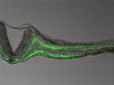
Photo: Above, an image of a tissue section of a wild quail embryo, injected with nanorods (red spot) used as probes to measure the rheology.